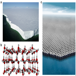
Peking University, Jan. 6, 2020: Water ices are ubiquitous in nature. The structure and growth of ice plays critical roles in an incredibly broad spectrum of materials science, tribology, biology, atmospheric science. As early as 1920s, Dennison, Bragg and Barnes studied the crystal structure of ice using X-ray diffractions, which represent the first attempts to learn about ice structures at molecular scale. For the following nearly 100 years, massive experiments and theoretical calculations have led to the discovery of 18 crystalline phases (three-dimensional phases), among which the hexagonal ice (Ice Ih) is the most common ice in the biosphere (Fig. 1a and b). However, whether the ice can exist at two dimensions (2D) has been under longstanding debate. At the monolayer limit, the 2D ice usually contains high-density unsaturated hydrogen bonds, and is thus unstable compared with the 3D case. Although 2D ice can be stabilized through the interaction with the substrate, such a "2D ice" largely relies on the structure and symmetry of the substrate, and cannot be considered as the genuine 2D ice.

Figure 1. a, The thick sheet floated on the Rose ocean in Antarctica. b, The crystal structure of Ice Ih. c, The 2D ice obtained in this work (rendered from experimental image).
Now, the teams led by Professor Jiang Ying, Professor Xu Limei and Professor Wang Enge of International Center for Quantum Materials (ICQM) of Peking University, in collaboration with Professor Xiao Cheng Zeng of Department of Physics of University of Nebraska-Lincoln, successfully grow a 2D bilayer hexagonal ice (named "2D ice I") and image the 2D ice growth at the edges with atomic resolution, through a combined study using scanning probe microscope (SPM), density functional theory (DFT) calculations and molecular dynamics (MD) simulations. This work is published in Nature on January 2, 2020.
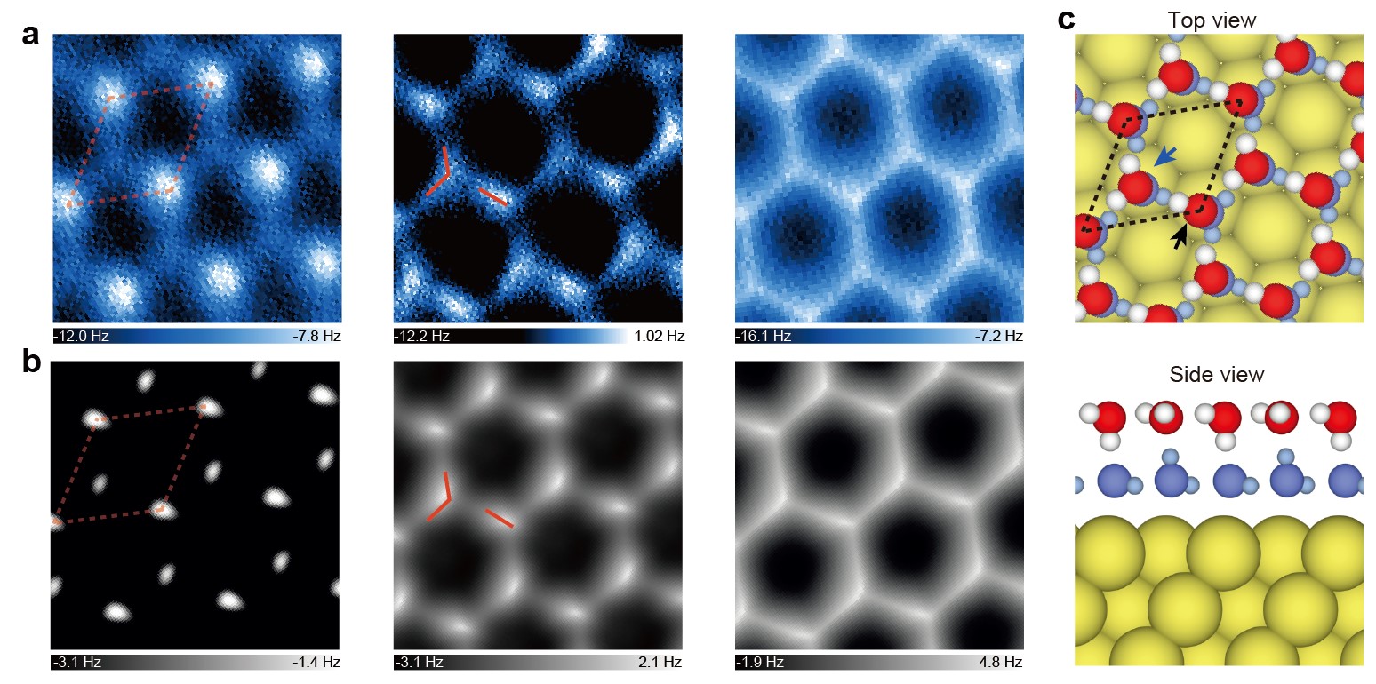
The key step is to choose the hydrophobic Au(111) surface as the substrate, which only weakly interacts with the water molecules. By carefully tuning the temperature and water pressure, the researchers were able to grow a single-crystal 2D ice fully wetting the surface (Fig. 1c). For the following, they used a qPlus-based atomic force microscopy (AFM) with CO-functionalized tip, which relies on the competition between the high-order electrostatic force and Pauli repulsion force, to image the 2D ice island (Fig. 2). The submolecular-resolution images reveal an interlocked bilayer-ice structure, consisting of two flat hexagonal water layers. In each water layer, half of the water molecules are lying flat while the other half are vertical with one O-H either upward or downward. The vertical water in one layer donates an H bond to the flat water in the other layer, leading to a fully saturated H-bonding structure. Such a stand-alone 2D ice was first predicted before by Koga et al. in 1997 using MD simulations, but direct imaging of its atomic structure has been lacking until now. Therefore, it is the first genuine 2D ice confirmed both by theory and experiment, i.e., named 2D ice I.

Figure 2. Detailed AFM characterization of the 2D bilayer ice and the corresponding structural model. a, b, Constant-height AFM imaging and simulated AFM images for different tip heights (the tip height decreases from left to right). c, Top and side views of the bilayer-ice structure on the Au(111) surface.
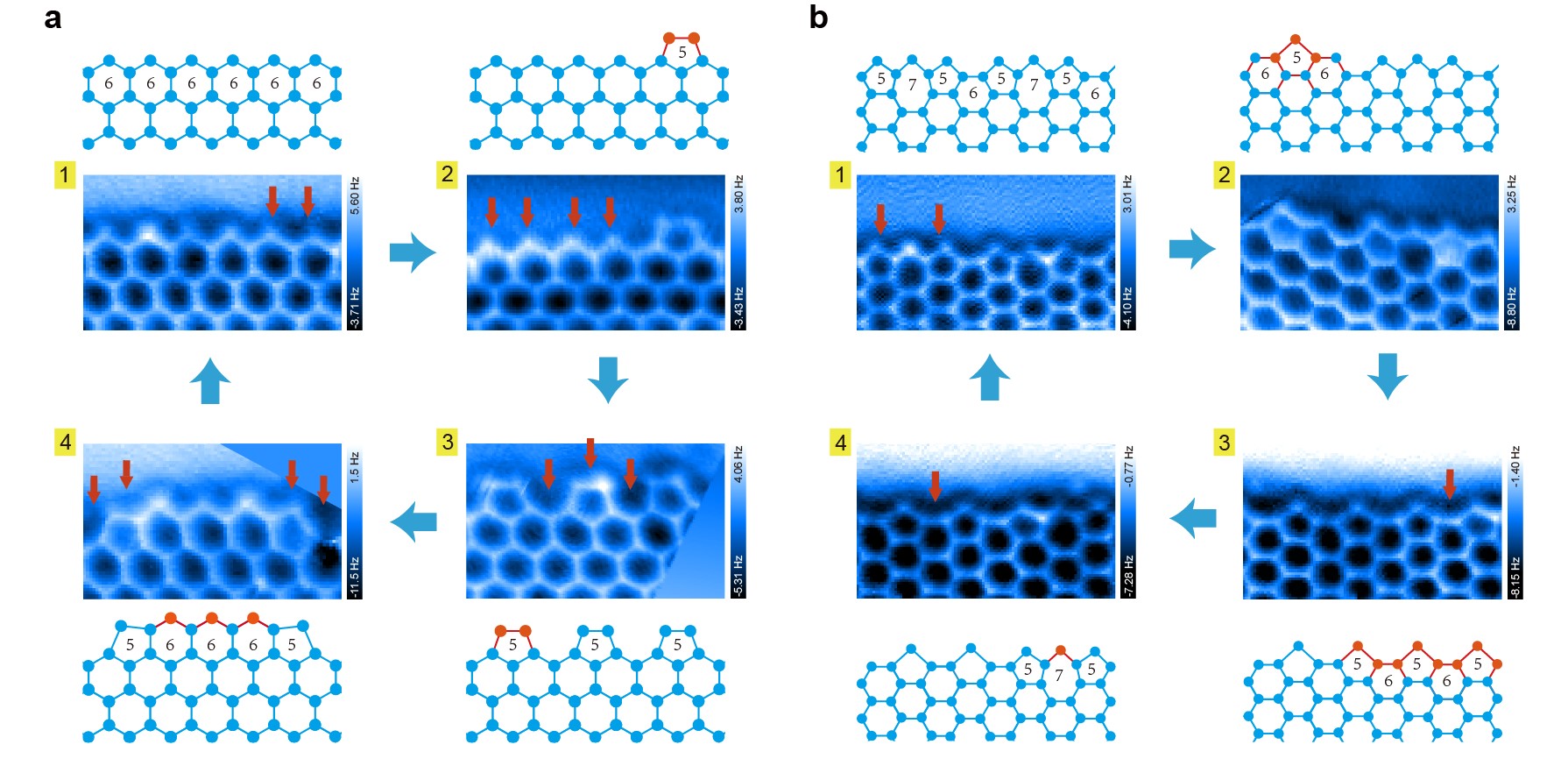
In order to understand the ice growth mechanism, the AFM was employed to image the edges of the 2D ice island. The disturbance of the CO-tip on the water can be reduced to a minimal level, thanks to the ultrahigh flexibility of the tip apex and the weak high-order electrostatic force between the tip and the water molecules. The weakly-perturbative nature of the probe overcomes the challenge of resolving highly fragile edge structures. Notably, the researchers deeply froze various metastable/intermediate edge structures involved in the ice growth, thus significantly prolonging their lifetimes to allow stable imaging. Those snapshots images were then used to reconstruct the ice growth process. They deduced very distinct ice growth behaviors, namely "collective bridging mode" for zigzag edge and "local seeding mode" for armchair edge, which are further corroborated by DFT and MD simulations. Therefore, the relative stability of different edge structures shows negligible dependence on the water spacing and the commensurability with the substrate, and such a growth behavior could be a generic phenomenon for 2D ice.

Figure 3. Proposed growing process for zigzag (a) and armchair (b) edges. Constant-height AFM images and corresponding ball-stick models of most stable (1) and metastable (2-4) of zigzag and armchair edges, respectively. The proposed process goes from 1 to 4.
This work confirms experimentally the existence of the first genuine 2D ice (2D ice I) and opens the door to explore other stand-alone 2D ice phases in nature. It is the first experiment to yield the atomic-scale information of ice growth in real space, which may renovate our conventional understanding of the formation and melting of low-dimensional ices on hydrophobic surfaces or under hydrophobic confinement. Furthermore, the techniques and methods developed in this work may be applicable to investigate the growth mechanism of a large family of 2D materials other than 2D ices, especially for those with multiple metastable/intermediate edge structures, thus opening up a new avenue of visualizing the structure and dynamics of low-dimensional matter at the cutting edges.
Dr. Ma Runze, Cao Duanyun, Tian Ye of International Center for Quantum Materials (ICQM) of Peking University and Zhu Chongqin of Department of Earth and Environmental Sciences of University of Pennsylvania are the co-first authors of this work. Professor Jiang Ying, Professor Wang Enge, Professor Xiao Cheng Zeng and Professor Xu Limei are the co-corresponding authors. This work received supports from National Natural Science Foundation of China, Ministry of Science and Technology of China, Chinese Academy of Sciences, and Collaborative Innovation Center of Quantum Matter.
Paper information: R. Ma, D. Cao, C. Zhu, Y. Tian, J. Peng, J. Guo, J. Chen, X.-Z. Li, J. S. Francisco, X. C. Zeng*, L.-M. Xu*, E.-G. Wang*, Y. Jiang*, "Atomic imaging of edge structure and growth of a two-dimensional hexagonal ice", Nature 577, 60–63 (2020).
Link: https://www.nature.com/articles/s41586-019-1853-4
Edited by: Huang Weijian
Source: International Center for Quantum Materials (ICQM) of Peking University